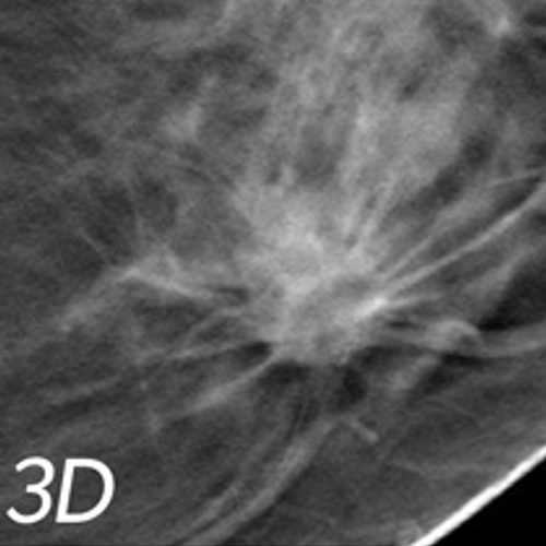
3D/TOMOSYNTHESIS DIGITAL MAMMOGRAPHY
Superior to 2D digital mammograms
Find cancer earlier than 2D mammograms alone and offer a 27% increase in cancer detection and a 40% increase in invasive cancer detection
Extraordinary technology allowing radiologists to look through breast tissue one millimeter at a time, seeing details inside the breast in a way never before possible
Provide less chance for cancer to hide behind overlapping tissue
Consist of multiple breast images taken in just seconds to produce a 3D image
what is digital mammography?
These images make it possible for a radiologist to gain a better understanding of your breast tissue and the confidence to reduce the need for follow-up imaging. By looking at the tissue in one-millimeter slices, the radiologist can provide a more confident assessment because now they can see breast tissue more clearly. With 3D mammography radiologists are able to find cancers earlier, with a 27% increase in cancer detection and a 40% increase in invasive cancer detection. Furthermore, 3D mammography significantly reduces callbacks by 20-40%.
If needed, non-surgical breast biopsy services, including ultrasound-guided and stereotactic biopsies, are available at our center.
Answers to questions you might have:
HOW DO I PREPARE FOR THE EXAM?
WHY 3D TOMOSYNTHESIS?
Doctors and scientists agree that early detection is the best defense against breast cancer, and successful treatment and survival rates are dramatically affected by it. If cancer is found early enough, before it has time to spread to lymph nodes, the five-year survival rate is almost 100 percent. Until now, the best way to identify breast cancer has been with digital mammography.
While digital mammography is still one of the best technologies available today, this is only a 2- dimensional picture of the breast. The breast is a 3-dimensional object composed of blood vessels, milk ducts and ligaments located at different heights and can overlap. When viewed as a 2-dimensional, flat image, these overlapping tissues can cause confusion and result in the leading reason as to why small breast cancers may be missed and normal tissue may appear abnormal, leading to unnecessary callbacks.
WHAT IS THE DIFFERENCE BETWEEN A SCREENING AND DIAGNOSTIC MAMMOGRAM?
A screening mammogram is your annual exam that is done every year. Sometimes the radiologist may ask you to come back for follow-up images, which is called a diagnostic mammogram, to rule out an unclear area in the breast or if there is a breast complaint that needs to be evaluated. 3D mammography may be used for both a screening mammogram and diagnostic mammogram, if you happened to be called back for this type of exam.
In a "conventional" 2D mammogram, there appears to be an area of concern that the doctor may want to further investigate with another mammogram or biopsy. Looking at the same breast tissue in 3D mammography imaging slices, the doctor can now see that the tissue is in fact normal breast tissue that was overlapping in the traditional mammogram, creating the illusion of an abnormal area. In this scenario, this patient likely avoided a callback for an additional mammogram, thanks to the 3D mammography exam technology.
Think of 3D mammogram technology like the pages of a book. If you look down at the cover, you cannot see all of the pages - but when you open it up, you can go through the entire book page by page to see everything between the covers.